Welcome!

Hello, and welcome to our site about Virus-Host Interactions. We are KGQMX, and we hope to help you understand more about viruses. Take a look at the side bar and learn fun, interesting facts at our fun facts section and also click on the quick links to learn more about the various topics available here.
Enjoy your time at our website! :D

Saturday, January 30, 2010

Virus Replication Cycle Part I

General Virus Replication Cycle

Below is the picture of a virus replication cycle.


From this picture, we can see that the virus undergoes 6 steps, mainly:

  • - Attachment
  • - Penetration
  • - Uncoating
  • - Replication & Expression
  • - Maturation
  • - And finally, the release of the Virus.

We'll cover in details, the different stages of virus replication at a later part.

This is what most of the virus will generally go through. However, there are also different types of the replication cycle.

  • 1. Lytic Cycle
  • 2. Lysogenic Cycle


(Image adapted from: http://www.prism.gatech.edu/~gh19/b1510/8lytic.jpg)

1. Firstly, attachment must occur before the virus goes into lytic or lysogenic cycle.
2. The phage DNA circularizes and enters lytic cycle or lysogenic cycle.

In the Lytic cycle:
3(i). New phage DNA and proteins are synthesized and assembeled into virions.
4(i).Cell lyses, releasing phage virions.
5(i). The cycle goes on again.

In the Lysogenic cycle:

3(ii). Phage DNA integrates within the bacterial chromosome by recombination, becoming a prophage.
4(ii). Lysogenic bacteria reproduces normally (undergoes many cell devisions).
5(ii). Occassionally, the prophage may seperate from the cell by another recombinant event, initiating a lytic cycle.

Now, we'll look into the Attachment stage.
1. Viral Attachment or Adsorption to the Host Cell

Adsorption (def: The attachment of one substance to the surface of another, eg, a virus attaching to a receptor on the surface of a susceptible host cell.) involves the binding of attachment sites on the viral surface with receptor sites on the host cell cytoplasmic membrane.

For a virus to infect a host cell, the cell must have specific receptors (ie, protein, glycoprotein or glycolipid) for the virus on its surface and also be capable of supporting viral replication. These host cell receptors are normal surface molecules involved in routine cellular function, but since a portion of a molecule on the viral surface resembles the chemical shape of the body's molecule that would normally bind to the receptor, the virus is able to attach to the host cell's surface.

An example is the Hepatitis B virus. The hepatitis B virus (HBV) adsorbs to IgA receptors on human cells. These receptors normally bind the antibody isotype IgA for transport across cells.


(Image adapted from: http://student.ccbcmd.edu/courses/bio141/lecguide/unit3/viruses/ev_adsorb.html)
The cell receptor site are specific. Virus binds to specific receptors on the host cell.
If the receptor on the host cell does not cater to the virus, then attachment will not happen.


(Image adapted from: http://www.goalfinder.com/product.asp?productid=151)
These are some of the receptors of certain host cells.

2. Penetration

Penetration into eukaryotic cells occurs in 3 ways:

  • Endocytosis
  • - Receptor-Mediated (enveloped virus)
  • - Clathrin (naked virus)
  • Fusion (enveloped virus)


(i) Endocytosis - Receptor-Mediated
Receptor Mediated Endocytosis - Image from academic.brooklyn.cuny.edu
(Image adapted from: http://academic.brooklyn.cuny.edu/biology/bio4fv/page/rectpr.htm)
The virus first binds to the receptor, then endocytosis happens.

(ii) Endocytosis - Clathrin


1. Attachment sites on the virus bind to corresponding receptors on the host cell membrane.


2. The virus begins to enter the host cell by endocytosis


3. The entire virus is placed in an endocytic vesicle.


4. The entire virus is placed in an endocytic vesicle.

(iii) Fusion


1. Attachment sites on the virus bind to corresponding receptors on the host cell membrane.


2. The viral envelope fuses with host cell's cytoplasmic membrane and nucleocapsid enters the host cell.


3. The viral envelope fuses with host cell's cytoplasmic membrane and nucleocapsid enters the host cell.

Virus Replication Cycle Part II

3. UNCOATING
Uncoating is a process that viral capsid is degraded by viral enzymes or host enzymes (for e.g. lysozymes).


Uncoating in general refers to the events that expose the viral genome to the host cellular machinery and sets the stage for the viral genome to express its functions required for the replication.

This stage occurs simultaneously with or rapidly after penetration. In order to express the viral genome to the cell organelles, it is necessary that the virion coat be removed partially or completely. Therefore, once virions are in the cytoplasm, they are generally uncoated to some extent by a variety of processes, including simple dissociation and/or enzyme-mediated partial degradation of the particles, to release the viral genome as a naked nucleic acid or as a nucleoprotein complex.

The steps involved in the process of disintegration of the protein coat or capsid to release its genome into the cell is called as uncoating. Uncoating may be achieved by the complete or partial removal of the capsid.


Penetration and endosomal uncoating

Uncoating of adenovirus and influenza virus.
Uncoating of HIV virus and paramyxoviridae virus.

4. REPLICATION
Replication involves assembly of viral proteins and genetic materials produced in the host cell.

The viral genome directs the host cell's metabolic machinery (ribosomes, tRNA, nutrients, energy, enzymes, etc.) to synthesize viral enzymes and viral parts. The viral genome has to both replicate itself and become transcribed into viral mRNA molecules. The viral mRNA can then be translated by the host cell's ribosomes into viral structural components and enzymes need for replication and assembly of the virus.Viruses can store their genetic information in six different types of nucleic acid which are named based on how that nucleic acid eventually becomes transcribed to the viral mRNA:

As the host cell's ribosomes attach to the viral mRNA molecules, the mRNAs are translated into viral structural proteins and viral enzymes. During the early phase of replication, proteins needed for the replication of the viral genome are made and the genome makes thousands of replicas of itself. During the late phase of replication, viral structural proteins (capsid and matrix proteins, envelope glycoproteins, etc.) and the enzymes involved in maturation are produced. Some viruses translate mRNA molecules that are transcripts of several genes into one or more large polyproteins. These polyproteins are subsequently cut into individual functional proteins by viral enzymes called proteases. Other viruses produce monocistronic mRNA molecules, each coding for a separate functional protein.

In the case of most RNA viruses, replication and assembly occurs in the host cell's cytoplasm. With DNA viruses, most replication and assembly occurs in the nucleus of the host cell. The viral genome enters the nucleus of the host cell and here is transcribed into viral mRNA. The viral mRNA molecules then leave the nucleus through the pores in the nuclear membrane and are translated into viral proteins by the host cell's ribosomes in the cytoplasm. Most of these viral proteins then re-enter the nucleus where the virus assembles around the replicated genomes.
Also during replication, viral envelope proteins and glycoproteins coded by the viral genome are incorporated into the host cell's cytoplasmic membrane (see Fig. 11A and Fig. 11B) or nuclear membrane.

5. MATURATION
Maturation is the assembly of protein capsid.

During maturation, the capsid is assembled around the viral genome (A complete set of genes).


6. RELEASE
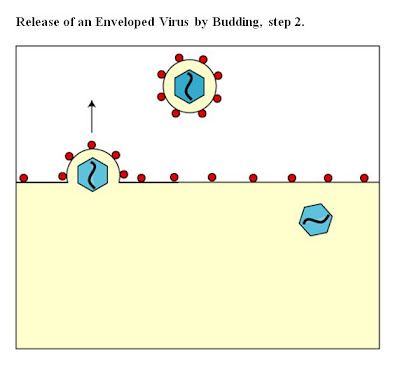
Viruses may escape from the host cell by causing cell rupture (lysis). Enveloped viruses (e.g., HIV) typically "bud" from the host cell.
During the budding process, a virus acquires the phospholipid envelope containing the embedded viral glycoproteins.

a. Naked viruses
Naked viruses are predominantly released by host cell lysis. While some viruses are cytolytic and lyse the host cell more or less directly, in many cases it is the body's immune defenses that lyse the infected cell.
b. Enveloped viruses
With enveloped viruses, the host cell may or may not be lysed. The viruses obtain their envelopes from host cell membranes by budding. As mentioned above, prior to budding, viral proteins and glycoproteins are incorporated into the host cell's membranes. During budding the host cell membrane with incorporated viral proteins and glycoproteins evaginates and pinches off to form the viral envelope. Budding occurs either at the outer cytoplasmic membrane, the nuclear membrane, or at the membranes of the Golgi apparatus
Viruses obtaining their envelope from the cytoplasmic membrane are released during the budding process.

Viruses obtaining their envelopes from the membranes of the nucleus, the endoplasmic reticulum, or the Golgi apparatus are then released by exocytosis(During exocytosis, a cell releases waste products or specific secretion products by the fusion of a vesicle with the cytoplasmic membrane) via transport vesicles.


Some viruses, capable of causing cell fusion, may be transported from one cell to adjacent cells without being released, that is, they are transmitted by cell-to-cell contact whereby an infected cell fuses with an uninfected cell.

Virus Life Cycle

Growth Curve


Attachment stage

Viruses bind to the receptor but have not get into the host yet.

Latent/ eclipse period

There is a fall in the number of viruses because no infectious particles are present during the replication process. Viruses will not be detected until it is being released.

Virus burst

Number of viruses went up because new progeny of viruses are assembled and released.

Classification of Viruses

Introduction

Virus classification involves naming and placing viruses into a taxonomic system. It is a subject of ongoing debate and proposals, largely due to the pseudo-living nature of viruses, which are not yet definitively living or non-living. As such, they do not fit neatly into the established biological classification system in place for cellular organisms.


Virus classification is based mainly on phenotypic characteristics, including morphology, nucleic acid type, mode of replication, host organisms, and the type of disease they cause. And also the chemical composition, the site of capsid assembly, the site of envelopment in enveloped viruses, the configuration of the nucleic acid and whether the genome is monopartite (meaning all genes are found within one segment of nucleic acid) or multipartite.

The genomic RNA strand of single-stranded RNA viruses is called sense (positive sense, plus sense) in orientation if it can serve as mRNA, and antisense (negative sense, minus sense) if a complementary strand synthesized by a viral RNA transcriptase serves as mRNA.


A combination of two main schemes is currently in widespread use for the classification of viruses. David Baltimore, a Nobel Prize-winning biologist, devised the Baltimore classification system, which places viruses into one of seven groups. These groups are designated by Roman numerals and separate viruses based on their mode of replication, and genome type.
Accompanying this broad method of classification are specific naming conventions and further classification guidelines set out by the International Committee on Taxonomy of Viruses.


Virosphere 2005


ICTV Classification


The International Committee on Taxonomy of Viruses began to devise and implement rules for the naming and classification of viruses early in the 1990s, an effort that continues to the present day. The ICTV is the only body charged by the International Union of Microbiological Societies (IUMS) with the task of developing, refining, and maintaining a universal virus taxonomy. The system shares many features with the classification system of cellular organisms, such as taxon structure. Viral classification starts at the level of order and follows as thus, with the taxon suffixes given in italics:


Order (-virales) - An order is a group of families sharing certain common characters.

Family (-viridae) - A family is a group of genera, whether or not these are organized into subfamilies, sharing certain common characters.

Subfamily (-virinae) - A subfamily is a group of genera sharing certain common characters. The taxon shall be used only when it is needed to solve a complex hierarchical problem.

Genus (-virus) - A virus genus is a group of related species that share some significant properties and often only differ in host range and virulence. Approval of a new genus must be accompanied by the approval of a type species.

Species - A species name shall consist of as few words as practicable but must not consist only of a host name and the word virus. A species name must provide an appropriately unambiguous identification of the species.


So far, six orders have been established by the ICTV: the Caudovirales, Herpesvirales, Mononegavirales, Nidovirales, Picornavirales, and Tymovirales.
These orders span viruses with varying host ranges.


- Caudovirales are tailed dsDNA (group I) bacteriophages
- Herpesvirales contains large eukaryotic dsDNA viruses
- Mononegavirales includes non-segmented (-) strand ssRNA (Group V) plant and animal viruses
- Nidovirales is composed of (+) strand ssRNA (Group IV) viruses with vertebrate hosts
- Picornavirales contains small (+) strand ssRNA viruses that infect a variety of plant, insect, and animal hosts
- Tymovirales contains monopartite ssRNA viruses that infect plants.

The establishment of an order is based on the inference that the virus families contained within a single order have most likely evolved from a common ancestor. The majority of virus families remain unplaced. Currently (2009) 6 orders, 87 families, 19 subfamilies, 348 genera, and 2,288 species of virus have been defined.


Baltimore Classification

Baltimore classification is a classification system that places viruses into one of seven groups depending on a combination of their nucleic acid (DNA or RNA), strandedness (single-stranded or double-stranded), Sense, and method of replication.


Picture for better understanding.


Classifying viruses according to their genome means that those in a given category will all behave in a similar fashion, offering some indication of how to proceed with further research.
Viruses can be placed in one of the seven following groups:


I: dsDNA viruses - viruses possess double-stranded DNA.(e.g. Adenoviruses, Herpesviruses, Poxviruses)

II: ssDNA viruses (+)sense DNA - viruses possess single-stranded DNA.
(e.g. Parvoviruses)

III: dsRNA viruses - viruses possess double-stranded RNA genomes, which are always segmented (e.g. Reoviruses)

IV: (+)ssRNA viruses (+)sense RNA - viruses possess positive-sense single-stranded RNA genomes. Many well known viruses are found in this group.
(e.g. picornaviruses, SARS virus, hepatitis C virus, yellow fever virus, and rubella virus.)

V: (−)ssRNA viruses (−)sense RNA - viruses possess negative-sense single-stranded RNA genomes.(e.g. Orthomyxoviruses, Rhabdoviruses, Ebola and Marburg viruses, influenza virus, measles, mumps and rabies)

VI: ssRNA-RT viruses (+)sense RNA with DNA intermediate in life-cycle - viruses possess single-stranded RNA genomes and replicate using reverse transcriptase. (e.g. Retroviruses)

VII: dsDNA-RT viruses - viruses possess double-stranded DNA genomes and replicate using reverse transcriptase.(e.g. Hepadnaviruses, Hepatitis B)


References:
http://en.wikipedia.org/wiki/Virus_classification
http://en.wikipedia.org/wiki/Baltimore_classification
http://www.ncbi.nlm.nih.gov/bookshelf/br.fcgi?book=mmed&part=A2252

Portal of Entry and Portal of Exit


Virus can enter into our body at many different sites and these are called portals of entry.

Skin

Skin when intact is usually a barrier to most pathogen as it has a tough outer layer of cornified cells. However, our skin is frequently breached by trauma or by inoculation (e.g. Cuts and insect bites). Virus inoculation by injection or transfusion is now common as a result both of medical procedures and of social practices such as sharing needles by intravenous drug users. Usually hepatitis B and hepatitis C viruses are usually transmitted in this way. They may also be transferred by minor “surgical” procedures like tattooing, dentistry and ear piercing. Furthermore, some pathogens can enter our body via our hair follicles, sweat glands, cuts and bruises.


Respiratory

Respiratory is one of the most common route of viral infection. As the average human adult breathes in about 600 L of air every hour; small suspended particles ( <>


Alimentary Tract

Although the surface of the alimentary tract is potentially exposed to a great number and variety of viruses, the harsh conditions in the stomach and duodenum protect it from many viruses. For instance, viruses that have a lipid-containing envelope are usually inactivated by the acid, bile salts and enzymes that occur in the stomach and duodenum. Infection via the gut, therefore, is due to viruses that resist these chemicals. These viruses multiply in the cells of the small intestine and are excreted in the feces (Table48-3). Such viruses usually resist environmental conditions and may cause water- and food-borne epidemics.


Placenta

Placenta transfer of virus from parent to offspring, and may occur via the ovum, across the placenta, during birth, or via the mother's milk. Viruses that cross the placenta include rubella virus and cytomegaloviruses, which may cause congenital defects or severe neonatal disease, and HIV. It can also result in spontaneous abortion, birth defect and premature birth.

In the end viruses escape from the body via the same surfaces, often but not necessarily by the route used as a portal of entry. Usually they leave our bodies via secretion ( tears, saliva, vaginal, semen etc. ) or excretion ( faeces, urine )


Virulence Factors

Viruses are pathogens that are able to cause disease. The ability of this pathogenicity is termed as virulence. Meaning, the more the pathogen is able to cause a disease, the more virulent it is.

Factors that enable pathogens to be more virulent mainly consists of the pathogen adhering and colonization in the host, evading the host’s immune system, entering and exiting host cells, and obtaining nutrition from the host. With these factors, many of the host’s functions are inhibited, and usually, the host will die.

Below is a comparison between Bacterial Virulence and Viral Virulence.


Click to enlarge!

I’m focusing on viral virulence, but if you want to know more about bacterial virulence, such as what adhesion, extracellular enzymes, toxins, anti-phagocytic factors are, and what they do to infect and replicate in the host, check out this page 2 of this pdf.

Adhesion
Viruses have to attach to surface proteins of cells before infecting them.

Host Evasion
Some viruses are able to “hide” within host cells in order to avoid the host’s immune system.

Latency
Viral latency refers to when viruses lie dormant in the cell. This can be seen by the lysogenic part of a virus life cycle. The virus’s genetic material may combine with the host cell’s DNA and remain there indefinitely, not replicating or infecting other cells. This allows them to reactivate and begin producing large amounts of viral progeny (replicated virus) without the host being infected by new outside virus.

High Mutability
Some viruses are able to mutate to evade the host cell’s immune system. The immune system recognizes foreign particles(in this case, the virus) due to antigens on the surface of the virus. Before the immune system can act on the virus with antibodies, the virus change their surface antigens to avoid recognition from the body. This is called antigenic variation.

So you’ve seen the virulence factors, now, you must be a little worried about getting infected by the many different kinds of deadly viruses you’ve heard of, such as HIV, mumps virus, and Ebola virus. Well, fret not, as different viruses have different intensities of virulence.

The diagram below shows an example of the relative virulence of some viruses.

As you can see, the influenza virus is one of the most virulent viruses among humans. Influenza virus easily infects and cause diseases in the body, known commonly as the flu.

There are five stages of infectious disease, which are the order in which diseases often occur in. They are:

1. Incubation
2. Prodormal
3. Illness
4. Decline
5. Convalescence
Image from here


Incubation
During the incubation period, the pathogen infects and replicates rapidly in the host. There are no signs or symptoms during this period. However, the host is able to spread the disease to another host. Some incubation periods can range from less than a day for Salmonella food-borne infection, to up to 8 years for HIV!


Prodormal
The prodormal period signals to the host that there is an impending illness. Some symptoms and signs appear. (Not all diseases have the prodormal stage)


Illness
Also called the invasive phase, this is when the most severe symptoms and signs pop up and are very clear. The acme is the peak of the disease symptoms. If the host’s immune system is unsuccessful in overcoming the pathogens during the invasive/illness phase, the host will die. Otherwise, it will go on to the next stage.


Decline
During the decline phase, symptoms and signs decrease, as the person gets better and the infection is brought under control.


Convalescence
There are no signs or symptoms in the convalescence period. Cells in the host repair damage caused by the infection, and everything goes back to normal.


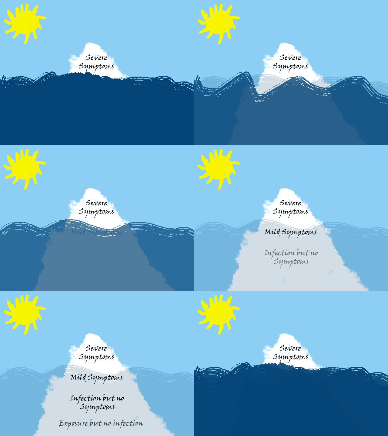
Something else related to the stages of infectious disease is the iceberg concept of infection. What is it? You’ve heard of the expression tip of the iceberg, well, the iceberg concept of infection is just like that, look at the image below.


Photobucket


I’m not that good at drawing on the computer, or drawing at all, so pardon the bad graphics and animation. The white mountain-shaped thing is the iceberg, and the blue moving things is the sea. Like the phrase “the tip of the iceberg” implies, when people realize that they have been infected, it is usually when they have severe symptoms. They do not realize the initial stages of infection before that, in the iceberg analogy, below the surface of the sea. The exposure, infection and mild symptoms usually go unnoticed.


Viruses are scary aren’t they? What frightens me the most is that they aren’t even living organisms; they behave and act like microorganisms such as parasitic bacteria, but do not have cells to divide. Virus infection also cannot be easily defeated by antibiotics like bacteria. The infection must be fought off by the body’s immune system or other drugs. There are so many different types of viruses and with the high ability to constantly mutate, it is pretty impossible to come up with new vaccines all the time. A widely known example is the human immunodeficiency virus (HIV); it has caused many deaths and long term suffering through AIDS, and despite many years of finding a cure for it, scientists have failed. More than 25 million people have died from AIDS since 1981, and it was estimated that there were 33.4 million more living with it at the end of 2008 (Avert.org).


What comforts me a little, is that viruses cannot survive for long when they leave the human body, as they are acellular particles, unable to carry out even the most basic life processes. They are not made up of cells, they cannot reproduce on their own, and they have no metabolism. So how do viruses spread from person to person, from plant to plant, from animal to animal?
Well, in order to maintain life outside of the host (whom they infect and replicate), they need to be maintained in sites called reservoirs of infection.


The three types of reservoirs are… (See animation below)


Photobucket

Note that the virus does not replicate in the reservoirs, but merely use them as a means (vector) to infect hosts, where they replicate.

Animal Reservoirs
Zoonoses (also called zoonotic diseases) are diseases that spread from animal hosts to humans. Humans are susceptible to infection from the virus when:

- They have direct contact with animals
- They have direct contact with faeces
- They eat the animals
- Vectors such as mosquitoes, fleas and other insects transfer fluids from animal to human.

Humans are usually the dead-end host to zoonotic pathogens.

Human Reservoirs
Some human virus reservoirs may appear to be healthy and asymtomatic, but these carriers actually are prone to infect others. The virus simply lies dormant in them, it may be that they do not have the protein receptor for the virus to attach to their cells, and thus preventing the virus from infecting them, or, that the human reservoir’s immune system is able to fight off the illness. Transfer of virus from human to human may be through contact or exchange of fluids.

Non-living Reservoirs
Soil, water and food are some examples of non-living reservoirs. Usually, the presence of viruses in these places is due to contamination with faeces and urine.


Ah, there you go. Now you know about virus virulent factors, stages of infectious disease, the iceberg concept of infection, and the reservoirs that viruses reside in before they infect. I hope that you have now understood, and learned more about viruses!


- Kiat Yi